IMMUNOLOGY & MICROBIOLOGY DEPARTMENT
In this issue of the newsletter, we will be highlighting the research being carried out by
Immunology & Microbiology Department at DDI, led by Dr. Rasheed Ahmad.
RESEARCH UPDATE
Find out what is new within the Research Sector at DDI
In-situ Protein Detection “the Power Of In-cell Western Blotting”
Published on 24/01/2020
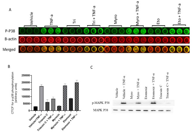
Western blot is a powerful technique originated in the laboratory of Harry Towbin at the Friedrich Miescher Institute in Basel, Switzerland in 1979. This sensitive immunological method identifies proteins using high affinity antigen-antibody interactions. This two-day long technique starts by isolating proteins from samples, which are then separated by size using slab gel electrophoresis. Separated proteins are then transferred to a suitable membrane that will serve as a carrier for all subsequent immunochemical reactions and imaging. With several steps involved, western blotting requires a significant investment in terms of equipment, training and reagents. With only few samples processed at a given time, it became clear that this method can be the bottleneck of any research workflow. As a solution, in-cell western blotting was introduced. Using the hybrid accuracy of protein quantification of western blotting, and the high-throughput of ELISA, in-cell western blot can be performed directly in 96-well microplates without the need to isolate the proteins from the sample or transfer this protein into any membrane. In a simpler manner, cells are grown on a sterile microplate, treated as required and directly fixed and permeabilized in situ. Then, usual techniques of labeling with a specific primary antibody followed by a fluorescent-conjugated secondary antibody are performed. Conjugates in the near-infrared (NIR) spectrum are commonly used to reduce auto-fluorescence and noise typically associated with tissue culture plastics. In addition, other florescence dyes can be used according to the imaging equipment capacity. This multiplex capacity allows for the assessment of multiple proteins of interest in a single well, which is useful for looking at both total and phosphorylated versions of a protein in a well, or to detect an in-well standard to allow for quantification across a plate (Figure 1).

Figure 1 Representative image of an in-cell western blot. Cells were cultured in a 96-well plate and treated with different lipid enzymes inhibitors. (A) Multicolor in-cell western blot. (B) Calculated total fluorescence of in-cell western blot data. (C) Traditional western blot.
REFERENCES
Lee, Y. S., Wollam, J. and Olefsky, J. M., An Integrated View of Immunometabolism. Cell 2018. 172: 22-40.
Epigenetic landscape priming by high fat dietary nutrients and adipokines plays a major role in metabolic inflammation and diabetes
Yang, Y., Zeng, C., Lu, X., Song, Y., Nie, J., Ran, R., Zhang, Z., He, C., Zhang, W. and Liu, S. M., 5-Hydroxymethylcytosines in Circulating Cell-Free DNA Reveal Vascular Complications of Type 2 Diabetes. Clin Chem 2019. 65: 1414-1425.


